
Kit de tinción de células muertas fijables Live or Dead™ *Fluorescencia azul con excitación de 405 nm * proporciona todos los componentes esenciales con un protocolo de etiquetado celular optimizado.
Descripción
Kit de tinción de células muertas fijables Live or Dead™ *Fluorescencia azul con excitación de 405 nm*
Nuestros kits de tinción de células muertas fijables Live or Dead™ son un conjunto de herramientas para etiquetar células para investigaciones microscópicas de fluorescencia de funciones celulares. El etiquetado eficaz de las células proporciona un método poderoso para estudiar eventos celulares en un contexto espacial y temporal.
Este kit en particular está diseñado para etiquetar uniformemente células de mamíferos fijadas en fluorescencia azul para aplicaciones de citometría de flujo con excitación con láser violeta. El kit utiliza un tinte fluorescente azul patentado que es más fluorescente al unirse a los componentes celulares y se excita bien con el láser violeta (excitación de 405 nm) hasta alcanzar una fluorescencia de 460 nm.
El kit proporciona todos los componentes esenciales con un protocolo de etiquetado celular optimizado. Es una herramienta excelente para preservar imágenes fluorescentes de células particulares y también puede usarse para aplicaciones de citometría de flujo de fluorescencia.
Nombre en Ingles: Live or Dead™ Fixable Dead Cell Staining Kit *Blue Fluorescence with 405 nm Excitation*
| Catalogo | Producto | Presentación |
|---|---|---|
| AAT-22500 | Kit de tinción de células muertas fijable Live or Dead™ * Fluorescencia azul con excitación de 405 nm* | 200 pruebas |


Importante: Solo para uso en investigación (RUO). Almacenamiento a <-15 °C. Minimizar exposición a la luz. Producto se entrega con geles.
Componentes
| Component A: Stain It™ Violet 450 | 1 vial |
| Component B: DMSO | 1 vial (200 µL) |
Plataforma
Citómetro de Flujo
| Excitación | 405 nm laser |
| Emisión | 450/40 nm filtro |
| Epecificaciones Instrumento | Canal Pacific Blue |
Microscopio de Fluorescencia
| Excitación | 410 nm |
| Emisión | 450 nm |
| Placa Recomendada | Pared negra / fondo claro |
Espectro
Abrir en Advanced Spectrum Viewer

Propiedades Espectrales
| Excitación (nm) | 406 |
| Emisión (nm) | 445 |
Imagen

Figura 1. Detección de la viabilidad de las células Jurkat mediante los kits de tinción de células muertas reparables Live or Dead™ (n.º de cat. 22500). Las células Jurkat se trataron y tiñeron con Stain It™ Violet 450 y luego se fijaron en formaldehído al 3,7 % y se analizaron mediante citometría de flujo. Las células vivas (rojo), tratadas con estaurosporina (verde) y tratadas térmicamente (azul) se distinguieron con el canal Pacific Blue.
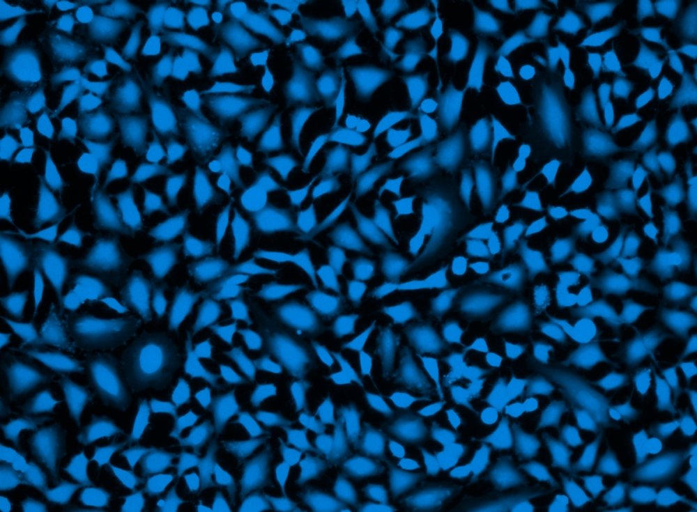
Figura 2. Imagen de células Hela fijadas con formaldehído y teñidas con el kit de tinción de células muertas fijable Live or Dead™ *Fluorescencia azul con excitación de 405 nm* en una placa Costa de 96 pocillos con pared negra/fondo transparente.
Formatos Alternativos
Productos Relacionados
Bibliografia
Autophagy proteins are not universally required for phagosome maturation
Authors: Cemma, Marija and Grinstein, Sergio and Brumell, John H
Journal: Autophagy (2016): 1440–1446
Differential detection of tumor cells using a combination of cell rolling, multivalent binding, and multiple antibodies
Authors: Myung, Ja Hye and Gajjar, Khyati A and Chen, Jihua and Molokie, Robert E and Hong, Seungpyo
Journal: Analytical chemistry (2014): 6088–6094
Versatile fabrication of nanoscale sol–gel bioactive glass particles for efficient bone tissue regeneration
Authors: Lei, Bo and Chen, Xiaofeng and Han, Xue and Zhou, Jiaan
Journal: Journal of Materials Chemistry (2012): 16906–16913
Referencias
Ver todas las 26 referencias: Citation Explorer
Requirements, features, and performance of high content screening platforms
Authors: Gough AH, Johnston PA.
Journal: Methods Mol Biol (2007): 41
A pharmaceutical company user’s perspective on the potential of high content screening in drug discovery
Authors: Hoffman AF, Garippa RJ.
Journal: Methods Mol Biol (2007): 19
Optimizing the integration of immunoreagents and fluorescent probes for multiplexed high content screening assays
Authors: Giuliano KA., undefined
Journal: Methods Mol Biol (2007): 189
Past, present, and future of high content screening and the field of cellomics
Authors: Taylor DL., undefined
Journal: Methods Mol Biol (2007): 3
High-content fluorescence-based screening for epigenetic modulators
Authors: Martinez ED, Dull AB, Beutler JA, Hager GL.
Journal: Methods Enzymol (2006): 21
Application of laser-scanning fluorescence microplate cytometry in high content screening
Authors: Bowen WP, Wylie PG.
Journal: Assay Drug Dev Technol (2006): 209
High-content screening of known G protein-coupled receptors by arrestin translocation
Authors: Hudson CC, Oakley RH, Sjaastad MD, Loomis CR.
Journal: Methods Enzymol (2006): 63
Evaluation of a high-content screening fluorescence-based assay analyzing the pharmacological modulation of lipid homeostasis in human macrophages
Authors: Werner T, Liebisch G, Gr and l M, Schmitz G.
Journal: Cytometry A (2006): 200
Automated high content screening for phosphoinositide 3 kinase inhibition using an AKT 1 redistribution assay
Authors: Wolff M, Haasen D, Merk S, Kroner M, Maier U, Bordel S, Wiedenmann J, Nienhaus GU, Valler M, Heilker R.
Journal: Comb Chem High Throughput Screen (2006): 339
High concordance of drug-induced human hepatotoxicity with in vitro cytotoxicity measured in a novel cell-based model using high content screening
Authors: O’Brien P J, Irwin W, Diaz D, Howard-Cofield E, Krejsa CM, Slaughter MR, Gao B, Kaludercic N, Angeline A, Bernardi P, Brain P, Hougham C.
Journal: Arch Toxicol (2006): 580
Application Notes
A New Red Fluorescent & Robust Screen Quest™ Rhod-4™ Ca2+Indicator for Screening GPCR & Ca2+ Channel Targets
A New Robust No-Wash FLIPR Calcium Assay Kit for Screening GPCR and Calcium Channel Targets
A Novel NO Wash Probeniceid-Free Calcium Assay for Functional Analysis of GPCR and Calcium Channel Targets
Abbreviation of Common Chemical Compounds Related to Peptides
Annexin V
FAQ
Are inflammasomes and caspase-1 related?
Question Are there any alternatives to BrdU (Bromodeoxyuridine)?
Are there upgraded trypan blue derivatives for cell viability testing?
Can 7-AAD be fixed?
Can 7-AAD stain live cells?
AssayWise
Nucleic Acid Detection, Quantification and Imaging
A practical guide for use of PE and APC in flow cytometry
Calbryte™ series now available
Fluorescent Phalloidin: A Practical Stain for Visualizing Actin Filaments
Intracellular pH Measurement with Dual Excitation Fluorescence Sensor BCFL