
El Ensayo de Viabilidad celular Cell Meter™ es útil para una variedad de estudios, incluida la adhesión celular, la quimiotaxis, la resistencia a múltiples fármacos, la viabilidad celular, la apoptosis y la citotoxicidad.
Descripción
Ensayo de Viabilidad celular Cell Meter™
Nuestros kits de ensayo Cell Meter™ son un conjunto de herramientas para monitorear la viabilidad celular. Hay una variedad de parámetros que se pueden usar para monitorear la viabilidad celular.
Este kit utiliza un tinte no fluorescente patentado que mejora la fluorescencia roja al entrar en las células vivas. El colorante es un compuesto hidrofóbico que penetra fácilmente en las células vivas intactas. La hidrólisis del sustrato no fluorescente por esterasas intracelulares genera un producto hidrofílico fuertemente fluorescente que se retiene bien en el citoplasma celular. La actividad de la esterasa es proporcional al número de células viables y, por lo tanto, está directamente relacionada con la intensidad de la fluorescencia del producto generado a partir de la hidrólisis catalizada por la esterasa del sustrato fluorogénico. Las células cultivadas en placas de paredes negras se pueden teñir y cuantificar en menos de dos horas.
El ensayo es más sólido que los ensayos basados en sal de tetrazolio o Alarmar Blue™. Se puede adaptar fácilmente para ensayos de alto rendimiento en una amplia variedad de plataformas de fluorescencia, como ensayos en microplaca, inmunocitoquímica y citometría de flujo. Es útil para una variedad de estudios, incluida la adhesión celular, la quimiotaxis, la resistencia a múltiples fármacos, la viabilidad celular, la apoptosis y la citotoxicidad.
El kit proporciona todos los componentes esenciales con un protocolo de etiquetado celular optimizado. Es adecuado para células proliferantes y no proliferativas, y se puede utilizar tanto para células en suspensión como adherentes. El indicador fluorogénico tiene propiedades espectrales compatibles con el juego de filtros Cy3/TRITC.
Nombre en Ingles: Cell Meter™ Cell Viability Assay Kit Red Fluorescence
| Catalogo | Producto | Presentación |
|---|---|---|
| AAT-22783 | Kit de ensayo de viabilidad celular Cell Meter™ Fluorescencia Roja | 200 pruebas |
Importante: Solo para uso en investigación (RUO).
Plataforma
Lector de Microplacas de Fluorescencia
| Excitación | 540 nm |
| Emisión | 590 nm |
| Cutoff | 570 nm |
| Placa recomendada | Pared negra /fondo claro |
| Especificaciones instrumento | Modo de lectura inferior |
Componentes
| Componente A: CytoCalcein™ Red AM | 2 viales, liofilizados |
| Componente B: DMSO | 1 vial (100 µL) |
| Componente C: Buffer de ensayo | 1 botella de 20ml |
Preparación de solución de stock
A menos que se indique lo contrario, todas las soluciones madre no utilizadas deben dividirse en alícuotas de un solo uso y almacenarse a -20 °C después de la preparación. Evite los ciclos repetidos de congelación y descongelación.
- Solución madre CytoCalcein™ Red AM:
Agregue 20 µL de DMSO (Componente B) en el vial de CytoCalcein™ Red AM (Componente A) y mezcle bien para preparar la solución madre de CytoCalcein™ Red AM. Nota: 20 µL de solución madre CytoCalcein™ Red AM es suficiente para una placa. Proteger de la luz. Para el almacenamiento, selle los tubos herméticamente. Nota: La solución madre de CytoCalcein™ Red sin usar puede dividirse en alícuotas y almacenarse a < -20oC durante un mes si los tubos están sellados herméticamente. Evite los ciclos repetidos de congelación y descongelación.
Preparación de solución de trabajo
Agregue todo el contenido (20 µL) de la solución madre de CytoCalcein™ Red AM en 10 mL de Tampón de ensayo (Componente C) y mezcle bien para preparar la solución de trabajo de CytoCalcein™ Red AM. Nota: Esta solución de trabajo CytoCalcein™ Red AM no es estable, úsela de inmediato. Nota: si las células, como las células CHO, contienen transportadores de aniones orgánicos que provocan la fuga del tinte fluorescente con el tiempo, se debe preparar una solución madre de probenecid y agregarla al tampón de carga a una concentración de trabajo final en el pozo de 1 -2,5 mM. Haga alícuotas y almacene la solución madre de probenecid no utilizada a ≤ -20 oC.
Para conocer guias sobre la preparación de muestras de células, visite: https://www.aatbio.com/resources/guides/cell-sample-preparation.html
Imagen
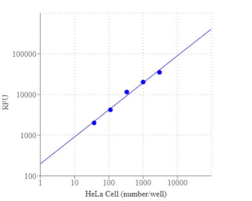
Figura 1. La respuesta del número de células HeLa se midió con el kit de ensayo de viabilidad celular Cell Meter™. Se sembraron células HeLa de 0 a 3000 células/pocillo/100 µl durante la noche en una placa de 96 pocillos Costar de pared negra/fondo transparente. Las células se incubaron con 100 µL/pocillo de solución de trabajo de colorante CytoCalcein™ Red durante 30 minutos a 37°C. La intensidad de la fluorescencia se midió a Ex/Em = 540/590 nm (corte = 570 nm) con el modo de lectura inferior utilizando Flexstation (de Molecular devices).
Formatos alternativos
Productos Relacionados
| Name | Excitation (nm) | Emission (nm) |
| Cell Meter™ Cell Viability Assay Kit *Green Fluorescence* | 494 | 514 |
Otros Productos
Cell Meter™ Intracellular Fluorimetric Hydrogen Peroxide Assay Kit *Green Fluorescence*
Cell Meter™ Intracellular Fluorimetric Hydrogen Peroxide Assay Kit *Blue Fluorescence*
Cell Meter™ Intracellular Fluorimetric Hydrogen Peroxide Assay Kit *Blue Fluorescence Optimized for Flow Cytometry*
Cell Meter™ Intracellular Fluorimetric Hydrogen Peroxide Assay Kit *Green Fluorescence Optimized for Flow Cytometry*
Cell Meter™ Mitochondrial Hydroxyl Radical Detection Kit *Red Fluorescence*
Cell Meter™ Fluorimetric Mitochondrial Superoxide Activity Assay Kit *Green Fluorescence*
Cell Meter™ Fluorimetric Intracellular Peroxynitrite Assay Kit *Green Fluorescence*
Cell Meter™ Fluorimetric Intracellular Peroxynitrite Assay Kit *Optimized for Flow Cytometry*
Cell Meter™ Fluorimetric Intracellular Nitric Oxide (NO) Activity Assay Kit *Orange Fluorescence Optimized for Microplate Reader*
Cell Meter™ Fluorimetric Intracellular Nitric Oxide (NO) Activity Assay Kit *Orange Fluorescence Optimized for Flow Cytometry*
Bibliografía
Ver todas las 18 bibliografias: Citation Explorer
Regulation of pancreatic stellate cell activation by Notch3
Authors: Song, Haiyan and Zhang, Yuxiang
Journal: BMC cancer (2018): 36
Functional imaging of neuronal activity of auditory cortex by using Cal-520 in anesthetized and awake mice
Authors: Li, Jingcheng and Zhang, Jianxiong and Wang, Meng and Pan, Junxia and Chen, Xiaowei and Liao, Xiang
Journal: Biomedical Optics Express (2017): 2599–2610
NINJ2–A novel regulator of endothelial inflammation and activation
Authors: Wang, Jingjing and Fa, Jingjing and Wang, Pengyun and Jia, Xinzhen and Peng, Huixin and Chen, Jing and Wang, Yifan and Wang, Chenhui and Chen, Qiuyun and Tu, Xin and others, undefined
Journal: Cellular Signalling (2017)
Influence of hypothermia and subsequent rewarming upon leukocyte-endothelial interactions and expression of Junctional-Adhesion-Molecules A and B
Authors: Bogert, Nicolai V and Werner, Isabella and Kornberger, Angela and Meybohm, Patrick and Moritz, Anton and Keller, Till and Stock, Ulrich A and Beiras-Fern, undefined and ez, Andres
Journal: Scientific reports (2016)
Inhibition of ABC transport proteins by oil sands process affected water
Authors: Alharbi, Hattan A and Saunders, David MV and Al-Mousa, Ahmed and Alcorn, Jane and Pereira, Alberto S and Martin, Jonathan W and Giesy, John P and Wiseman, Steve B
Journal: Aquatic Toxicology (2016): 81–88
Rapid generation of collagen-based microtissues to study cell–matrix interactions
Authors: Brett, Marie-Elena and Crampton, Alex and ra L , undefined and Wood, David K
Journal: Technology (2016): 1–8
Toxicokinetics and toxicodynamics of chlorpyrifos is altered in embryos of Japanese medaka exposed to oil sands process-affected water: evidence for inhibition of P-glycoprotein
Authors: Alharbi, Hattan A and Alcorn, Jane and Al-Mousa, Ahmed and Giesy, John P and Wiseman, Steve B
Journal: Journal of Applied Toxicology (2016)
Flexible Endoscopic Spray Application of Respiratory Epithelial Cells as Platform Technology to Apply Cells in Tubular Organs
Authors: Thiebes, Anja Lena and Reddemann, Manuel Armin and Palmer, Johannes and Kneer, Reinhold and Jockenhoevel, Stefan and Cornelissen, Christian Gabriel
Journal: Tissue Engineering Part C: Methods (2016): 322–331
Erythropoietin Stimulates Endothelial Progenitor Cells to Induce Endothelialization in an Aneurysm Neck After Coil Embolization by Modulating Vascular Endothelial Growth Factor
Authors: Liu, Peixi and Zhou, Yingjie and An, Qingzhu and Song, Yaying and Chen, Xi and Yang, Guo-Yuan and Zhu, Wei
Journal: MEDICINE (2016): 1–8
Spraying respiratory epithelial cells to coat tissue-engineered constructs
Authors: Thiebes, Anja Lena and Albers, Stefanie and Klopsch, Christian and Jockenhoevel, Stefan and Cornelissen, Christian G
Journal: BioResearch open access (2015): 278–287
Referencias
Ver todas las 84 referencias: Citation Explorer
Functional evidence that the self-renewal gene NANOG regulates esophageal squamous cancer development
Authors: Li, Deng and Xiang, Xiaocong and Yang, Fei and Xiao, Dongqin and Liu, Kang and Chen, Zhu and Zhang, Ruolan and Feng, Gang
Journal: Biochemical and Biophysical Research Communications (2017)
Localized functional chemical stimulation of TE 671 cells cultured on nanoporous membrane by calcein and acetylcholine
Authors: Zibek S, Stett A, Koltay P, Hu M, Zengerle R, Nisch W, Stelzle M.
Journal: Biophys J. (2006)
A vaccination and challenge model using calcein marked fish
Authors: Klesius PH, Evans JJ, Shoemaker CA, Pasnik DJ.
Journal: Fish Shellfish Immunol (2006): 20
Novel fluorescence assay using calcein-AM for the determination of human erythrocyte viability and aging
Authors: Bratosin D, Mitrofan L, Palii C, Estaquier J, Montreuil J.
Journal: Cytometry A (2005): 78
Cytotoxic effects of 100 reference compounds on Hep G2 and HeLa cells and of 60 compounds on ECC-1 and CHO cells. I mechanistic assays on ROS, glutathione depletion and calcein uptake
Authors: Schoonen WG, Westerink WM, de Roos JA, Debiton E.
Journal: Toxicol In Vitro (2005): 505
Calcein AM release-based cytotoxic cell assay for fish leucocytes
Authors: Iwanowicz LR, Densmore CL, Ottinger CA.
Journal: Fish Shellfish Immunol (2004): 127
Calcein-AM is a detector of intracellular oxidative activity
Authors: Uggeri J, Gatti R, Belletti S, Sc and roglio R, Corradini R, Rotoli BM, Orl and ini G., undefined
Journal: Histochem Cell Biol (2004): 499
Comparison of the usefulness of the MTT, ATP, and calcein assays to predict the potency of cytotoxic agents in various human cancer cell lines
Authors: Mueller H, Kassack MU, Wiese M.
Journal: J Biomol Screen (2004): 506
In vitro assay of mineralized-tissue formation on titanium using fluorescent staining with calcein blue
Authors: Goto T, Kajiwara H, Yoshinari M, Fukuhara E, Kobayashi S, Tanaka T.
Journal: Biomaterials (2003): 3885
The effects of calcium chloride and sodium chloride on the electroporation-mediated skin permeation of fluorescein isothiocyanate (FITC)-dextrans in vitro
Authors: Tokudome Y, Sugibayashi K.
Journal: Biol Pharm Bull (2003): 1508
Application notes
Annexin V
Calcein
Multi-Color Labeling and Functional Analysis of Live Cells Using Fluorescent Calcein AM Dyes
Multiplexing Cell Proliferation and Cytotoxicity Assays Using Calcein Red and CytoCalceins
Relative Brightness of Fluorescent Dyes
FAQ
Are there any alternatives to BrdU (Bromodeoxyuridine)?
Are there upgraded trypan blue derivatives for cell viability testing?
Is Calcein, AM toxic to cells?
What are common laser lines used in flow cytometry?
What does trypan blue do?